01 / Reading Guide
Reading Guide
Citation
Ellis, H., & Mahadevan, V. (2018). Clinical Anatomy: Applied Anatomy for Students and Junior Doctors (14th ed.). Wiley-Blackwell.
Chapter Summary
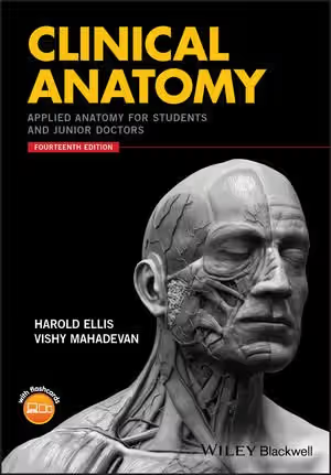
The fourteenth edition runs to about 500 pages in six parts, each organised by region and divided into short topics, followed by a glossary of eponyms and an index. Both the preface to this edition and the preface to the first edition are reprinted. Wiley describes the edition as revised to put more weight on clinical relevance, to add content for non-surgical trainees, to include illustrative clinical scenarios, to add many more CT, MRI and other images, some overlaid with anatomical drawings to show surface anatomy, and to expand the coverage of the nervous system. A companion website holds digital flashcards of the book's illustrations and photographs.
Part 1: The Thorax
The book opens with surface anatomy and surface markings of the chest, then the thoracic cage (vertebrae, ribs, costal cartilages, sternum, intercostal spaces) and the diaphragm, followed by the pleurae, the lower respiratory tract (trachea, bronchi, lungs), the mediastinum and pericardium, the heart, and the superior mediastinum with the thymus, oesophagus, thoracic duct and sympathetic trunk. It ends with practical sections on examining a chest radiograph and the radiographic appearance of the heart.
Part 2: The Abdomen and Pelvis
The longest part starts from vertebral levels and the surface markings of the viscera, moves to the abdominal wall (including the anatomy of abdominal incisions and the inguinal canal) and the peritoneal cavity with its fossae and subphrenic spaces, and then works through the gastrointestinal tract, its blood supply, portal venous system and lymphatic drainage, and its development and congenital abnormalities. Further topics cover the liver, biliary system, pancreas and spleen; the urinary tract with its embryology; the male genital organs; the bony pelvis, including obstetric measurements, variations in pelvic shape and sacral (caudal) anaesthesia; the pelvic floor and perineum; the female genital organs, including vaginal examination and embryology; the posterior abdominal wall; and a closing section on computed axial tomography.
Part 3: The Upper Limb
Surface anatomy comes first again, then the bones and joints from scapula to hand. Three "important zones" (axilla, cubital fossa, carpal tunnel) are given their own section, followed by the arteries, the brachial plexus and segmental cutaneous supply, the course of the principal nerves, compartments and compartment syndrome, the female breast with its lymphatic drainage, the anatomy of upper limb deformities, and the spaces of the hand.
Part 4: The Lower Limb
The lower limb mirrors the upper: surface anatomy (including bursae and limb measurement), bones and joints, the arches of the foot and the anatomy of walking, three important zones (femoral triangle, adductor canal, popliteal fossa), the arteries and veins, the lumbar and sacral plexuses and principal nerves, and compartments with compartment syndrome.
Part 5: The Head and Neck
This part moves from the surface anatomy and fascial compartments of the neck to the thyroid and parathyroid glands, the palate and the development of the face and palate with its congenital deformities, the tongue and floor of the mouth, the pharynx and the mechanism of swallowing, the larynx, the salivary glands, the major arteries and veins (including the dural venous sinuses), the lymph nodes of the neck, the cervical sympathetic trunk and the branchial system. It continues with the scalp, skull, paranasal sinuses, mandible and teeth, and ends with the vertebral column and intervertebral joints.
Part 6: The Nervous System
The final part, expanded in this edition, covers the brain (brainstem, cerebellum, diencephalon, pituitary, cerebral hemispheres, meninges, ventricular system and cerebrospinal fluid), the spinal cord and its membranes, each of the twelve cranial nerves in turn, the special senses (nose, ear and eye) and the autonomic nervous system.
Key Concepts
Parts 1–2: Thorax, abdomen and pelvis
- Surface markings: projecting deep organs onto landmarks on the body surface, the starting point of every part.
- Vertebral levels: using the spine as a reference for locating abdominal structures.
- Inguinal canal and abdominal incisions: abdominal wall anatomy treated from a surgical point of view.
- Portal venous system: the venous drainage of the gut to the liver.
- Radiological anatomy: reading a chest radiograph and CT sections.
Parts 3–4: The limbs
- Important zones: the axilla, cubital fossa and carpal tunnel, and the femoral triangle, adductor canal and popliteal fossa, as compact regions where key structures meet.
- Course and distribution of nerves: following each principal nerve to understand the effects of injury and the deformities that result.
- Compartment syndrome: the consequence of raised pressure within the fascial compartments of a limb.
Part 5: Head and neck
- Fascial compartments of the neck: the tissue planes that organise the anterior neck.
- Branchial system: the embryonic arches whose derivatives, cysts and fistulae appear in the neck.
- Developmental anomalies: the formation of the face, lips and palate and the congenital deformities that follow from it.
Part 6: The nervous system
- Cranial nerves: the twelve nerves treated individually, with the optic nerve linked to the visual pathway.
- Ventricular system and cerebrospinal fluid circulation: the fluid spaces of the brain.
- Autonomic nervous system: the sympathetic and parasympathetic systems.
Critical Analysis
Strengths
- Selected for clinical use. The subtitle and the publisher's description present the book as the anatomy that students and junior doctors need in a clinical setting. The table of contents bears this out with sections on abdominal incisions, chest radiographs, obstetric pelvic measurements, caudal anaesthesia, vaginal examination, compartment syndrome and limb deformities, topics that a purely descriptive text would not single out.
- Surface anatomy throughout. Every regional part opens with surface anatomy and surface markings, and this edition overlays anatomical drawings on images and photographs to show it.
- Embryology where it explains anomalies. Developmental sections appear where they account for congenital abnormalities: the gut, kidney and ureter, female genital tract, face and palate, and branchial system.
- A long-tested text. The book has reached its fourteenth edition and still carries the preface to the first, and Wiley names medical students, junior doctors and candidates for the Royal College of Surgeons examinations among its readers.
Limitations
- Not a complete anatomy course on its own. The regional coverage is selective, and the book contains no separate treatment of general anatomy, tissues or histology; students meeting anatomy for the first time may need an introductory or systemic text alongside it.
- A surgical emphasis. Both authors come from surgery (Wiley lists Ellis as Emeritus Professor of Surgery and Mahadevan as Professor of Surgical Anatomy at the Royal College of Surgeons of England), topics such as abdominal incisions reflect that, and the publisher notes that this edition adds more for non-surgical trainees. Students heading for medical rather than surgical specialties may find some emphases less relevant.
- Physiology is outside its scope. The nervous system part is anatomical; function and neuroscience need other books.
Where It Fits in the Course
In this anatomy course, Ellis and Mahadevan is the concise, clinically selected companion. Crash Course Anatomy and Physiology is the lighter revision guide, organised by region and aimed at exams, while Drake, Vogl and Mitchell's Gray's Anatomy for Students and Dalley and Agur's Moore's Clinically Oriented Anatomy are the full regional textbooks with extensive illustration, surface anatomy and imaging. A sensible pattern is to learn each region first from one of those and then use Ellis to see which details matter on the wards and in surgical examinations.
For reference beyond this, Gray's Anatomy: The Anatomical Basis of Clinical Practice provides the detailed account of structure, development and variation, and MacKinnon and Morris's Oxford Textbook of Functional Anatomy explains structure through function. Ellis is shorter than all of these and is best read alongside or after a main textbook rather than instead of one.
How to Use This Book
The publisher's audience is medical students, postgraduate trainees and junior doctors, including those preparing for Royal College of Surgeons examinations. School biology is enough to start, but readers will get more from it once a first pass through the regions has been made in dissection or a main textbook.
A practical plan:
- Follow the dissection or teaching order of your course. Each part stands on its own, so the parts can be read in the order a course teaches the regions.
- Start each region with its surface anatomy. Learn the surface markings first and test them on a classmate or on yourself.
- Prioritise the clinically labelled topics. In the thorax, the chest radiograph sections; in the abdomen, incisions, the inguinal canal, the peritoneal spaces and the gut's blood supply; in the limbs, the important zones, nerve injuries, compartments and deformities; in the head and neck, the thyroid, larynx and neck lymph nodes.
- Use the imaging. Read the CT, MRI and radiograph material with real images when possible, since the edition puts weight on them.
- Revise with the companion website. The flashcards of every illustration and photograph are suitable for quick self-testing before examinations.
- Optional on a first pass: the embryology subsections and the glossary of eponyms, which are useful for understanding anomalies and named signs later.