01 / Reading Guide
Reading Guide
Citation
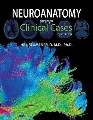
Blumenfeld, H. (2021). Neuroanatomy through Clinical Cases (3rd ed.). Sinauer Associates (Oxford University Press).
Chapter Summary
The book has nineteen chapters and a short epilogue. According to the publisher's description, each chapter has two halves: first background material, consisting of an overview of the relevant neuroanatomical structures and pathways and a brief discussion of related clinical disorders; then a set of clinical cases. Each case begins with a narrative of how the patient developed symptoms and what was found on neurological examination, and a series of questions asks the reader to deduce where the lesion is and what the diagnosis might be, before discussion and answers reveal the outcome. The publisher counts more than 100 real cases, accompanied by radiological images.
Chapters 1–4: Introduction to Clinical Case Presentations; Neuroanatomy Overview and Basic Definitions; The Neurologic Exam as a Lesson in Neuroanatomy; Introduction to Clinical Neuroradiology
The opening chapters set up the method: how cases are presented, the basic vocabulary of neuroanatomy, the neurological examination read as a test of specific anatomical systems, and an introduction to the neuroimaging used to see lesions. The publisher's companion website, neuroexam.com, supplies video demonstrations of the steps of the examination and notes which anatomical systems each step tests.
Chapters 5–9: Brain and Environs: Cranium, Ventricles, and Meninges; Corticospinal Tract and Other Motor Pathways; Somatosensory Pathways; Spinal Nerve Roots; Major Plexuses and Peripheral Nerves
These chapters move from the protective surroundings of the brain to the long motor and sensory pathways and then to the peripheral nervous system. The publisher's sample material for Chapter 6 gives a sense of the treatment: figures on the cortical motor and somatosensory areas, spinal cord cross-sections and arterial supply, the internal capsule and the descending motor pathways, a table contrasting upper and lower motor neuron lesion signs, and figures of weakness patterns (hemiparesis, monoparesis, paraparesis and quadriparesis among them) together with images of infarcts and other lesions.
Chapters 10–14: Cerebral Hemispheres and Vascular Supply; Visual System; Brainstem I: Surface Anatomy and Cranial Nerves; Brainstem II: Eye Movements and Pupillary Control; Brainstem III: Internal Structures and Vascular Supply
The middle of the book covers the cerebral hemispheres with their blood supply, the visual system, and the brainstem in three chapters: external anatomy and cranial nerves, eye movements and pupillary control, and internal structures with vascular supply. The brainstem is the only topic given three consecutive chapters.
Chapters 15–19 and Epilogue: Cerebellum; Basal Ganglia; Pituitary and Hypothalamus; Limbic System: Homeostasis, Olfaction, Memory and Emotion; Higher-Order Cerebral Function; Epilogue: A Simple Working Model of the Mind
The final chapters treat the cerebellum, the basal ganglia, the pituitary and hypothalamus, and the limbic system, and end with higher-order cerebral function. The epilogue offers a short working model of the mind.
Key Concepts
Chapters 1–4: Method and tools
- Lesion localisation: the book's organising skill, deducing the site of damage from the history and examination.
- The neurologic exam as applied anatomy: Chapter 3's title frames the examination as a lesson in neuroanatomy.
- Clinical neuroradiology: Chapter 4 introduces the imaging used to confirm where a lesion lies.
Chapters 5–9: Surroundings, pathways and periphery
- Cranium, ventricles and meninges: the coverings and fluid spaces of the brain (Chapter 5).
- Corticospinal tract and other motor pathways: the descending pathways whose damage produces the weakness patterns in Chapter 6.
- Somatosensory pathways: the ascending routes for bodily sensation (Chapter 7).
- Spinal nerve roots, plexuses and peripheral nerves: the transition from central to peripheral lesions (Chapters 8–9).
Chapters 10–19: Regions and systems
- Vascular supply of the hemispheres and brainstem: named in the titles of Chapters 10 and 14.
- Cranial nerves, eye movements and pupillary control: the brainstem material of Chapters 12–13.
- Cerebellum and basal ganglia: the motor-control structures of Chapters 15–16.
- Limbic system and higher-order cerebral function: Chapters 18–19.
Critical Analysis
Strengths
- Case-based design built into every chapter. The publisher describes a fixed pattern of background followed by cases that withhold the answer until the reader has reasoned about the lesion. For a student who learns by problem-solving, this gives regular practice at the skill a neurology clerkship tests.
- A sequence organised around pathways and regions. The contents place the motor and sensory pathways, roots and plexuses (Chapters 6–9) before the regional chapters, and give the brainstem three chapters of its own, so the structures are met in the order in which lesion localisation tends to use them.
- Examination and imaging taught alongside anatomy. Chapters 3 and 4 give the neurological examination and neuroradiology a place in the book rather than leaving them to a clinical course, and the publisher's companion site adds examination videos.
- Digital study material. The publisher's site lists, for the enhanced e-book, interactive figures and tables, review exercises and an interactive brief anatomical study guide per chapter, with Chapter 6 available as a sample; instructor resources include case conference slides and handouts.
Limitations
- Length. Catalogues give the third edition about 1,030 to 1,056 pages. The depth is welcome in a course built on cases, but it is heavy for a short introductory unit.
- A reviewer of the second edition found it text-heavy. A review on the Kenhub learning site judged that edition's pages to be dense with relatively few illustrations and its built-in review testing limited. The third edition is described as fully updated, with enhanced digital components, but the print layout itself is worth checking in a sample before buying.
- Not a quick-reference text. Because the teaching happens through narrative cases, a student who needs a compact summary of one pathway before an exam will find a short illustrated text easier to scan.
Where It Fits in the Course
Within the neuroscience course list, this is a clinically oriented neuroanatomy text. Snell's Clinical Neuroanatomy covers the nervous system in a systematic sequence of chapters (organisation, neurons, spinal cord, brainstem, cerebellum and so on) with clinical cases and review questions, so the two books can be used as alternatives: Snell's chapters are organised by structure and system, whereas Blumenfeld pairs every topic with cases. Shorter visual books, such as Neuroanatomy: Illustrated Colour Text and Neuroanatomy and Neuroscience at a Glance, give a quick map of the nervous system in double-page topics and suit a first pass or last-minute revision.
For the cellular and systems science behind the anatomy, the broader neuroscience textbooks in the course (Purves et al.'s Neuroscience, Bear, Connors and Paradiso's Neuroscience: Exploring the Brain, and the free Foundations of Neuroscience) are the companions. They explain how neurons signal and how sensory and motor systems work, and because the contents of Blumenfeld include no chapters on cellular neurophysiology, they fill that gap.
How to Use This Book
The publisher addresses first- and second-year medical students in a basic neuroanatomy, neurobiology or neuroscience course, and also advanced medical students, residents and students of health professions from physical therapy to dentistry. Because the publisher pitches it at students in a basic course, no earlier neuroanatomy is presupposed.
A practical plan: read Chapters 1–4 first, since they explain how the cases work and how the examination and imaging connect to the anatomy. Then follow the book in order through the pathways and regions, reading each background half, drawing the pathway involved, and attempting every case question on paper before reading the discussion. Chapters 6, 7, 12–14 and 15–16 repay the closest work for a course with a neurology emphasis. The epilogue and the final chapters on higher-order function can be read last or used for enrichment. Use neuroexam.com alongside Chapter 3, and the enhanced e-book's review exercises and study guides if they are available with your edition.