01 / Reading Guide
Reading Guide
Citation
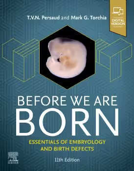
Torchia, M. G., & Persaud, T. V. N. (2024). Before We Are Born: Essentials of Embryology and Birth Defects (11th ed.). Elsevier.
Chapter Summary
The eleventh edition runs to about 350 pages in twenty chapters, followed by a bibliography and an appendix of answers to the clinically oriented questions. Elsevier describes it as based on the authors' longer textbook, The Developing Human, with coverage selected to give the essentials of normal and abnormal development, the causes of birth defects and the role of genes. According to the publisher, every chapter includes clinical cases, the book carries nearly 700 USMLE-style questions with answers and explanations, and purchasers have access to 18 narrated animations and an eBook. Terminology follows Terminologia Embryonica (2019). The chapters fall into three groups: a chronological account of development, a set of organ-system chapters, and two closing chapters on birth defects and on the molecular basis of development.
Chapters 1–2: Introduction to Human Development; Human Reproduction
The opening chapters introduce the field and its terminology and then cover human reproduction, the background needed before development begins.
Chapters 3–5: First Week; Second Week; Third Week of Human Development
Three chapters follow the embryo week by week through the earliest stages, from the first week after fertilisation to the third week.
Chapters 6–7: Fourth to Eighth Weeks of Human Development; Fetal Period
The embryonic period proper (weeks four to eight) is treated in one chapter, and the fetal period, from the end of the eighth week to birth, in the next.
Chapter 8: Placenta and Fetal Membranes
The structures that support the embryo and fetus close the chronological part of the book.
Chapters 9–10: Body Cavities, Mesenteries, and Diaphragm; Pharyngeal Apparatus, Face, and Neck
The systematic part begins with the division of the body cavities and the formation of the diaphragm, then the pharyngeal apparatus and the development of the face and neck.
Chapters 11–14: Respiratory System; Alimentary System; Urogenital System; Cardiovascular System
The internal organ systems, each followed from its origins through to its definitive form.
Chapters 15–18: Musculoskeletal System; Nervous System; Development of Eyes and Ears; Integumentary System
The remaining systems, including the special senses in a chapter of their own.
Chapter 19: Human Birth Defects
A chapter devoted to birth defects as a whole. The publisher notes an updated teratology section, the study of environmental causes of abnormal development.
Chapter 20: The Cellular and Molecular Basis of Development
The book ends, rather than begins, with the cellular and molecular mechanisms of development, on which the publisher says this edition adds new information.
Key Concepts
Chapters 1–8: Stages of development
- Embryonic and fetal periods: the first eight weeks, in which the main structures form, and the period of growth and maturation that follows.
- Week-by-week development: the sequence of events in the first three weeks, each given its own chapter.
- Placenta and fetal membranes: the structures that connect embryo and fetus with the mother.
Chapters 9–18: Organ systems
- Body cavities and diaphragm: how the single embryonic body cavity is divided.
- Pharyngeal apparatus: the embryonic structures from which much of the face and neck develop.
- Organogenesis: the development of each organ system from its origins to its adult form.
Chapters 19–20: Birth defects and mechanisms
- Teratology: the study of environmental factors that cause birth defects.
- Molecular basis of development: the genes and cellular signals that control embryonic development.
Critical Analysis
Strengths
- A short version of a standard text. The book derives from The Developing Human by the same authors and condenses it to about 350 pages, so students get the same framework and terminology in a length suited to a short embryology component.
- Clinical orientation. The publisher states that every chapter includes clinical cases linking development to practice, and the subtitle and Chapter 19 put birth defects at the centre rather than in an appendix.
- Extensive self-testing. Nearly 700 USMLE-style questions with full explanations, plus the clinically oriented questions answered in the appendix, give a large amount of practice for a book of this length.
- Visual resources. Elsevier highlights new colour photographs and diagnostic images (3D ultrasound, CT and MR), and the 18 narrated animations are aimed at key developmental concepts, many of which involve three-dimensional changes that are hard to follow on the page.
Limitations
- Molecular mechanisms come last. Chapter 20 places the cellular and molecular basis of development at the end; students whose course teaches signalling pathways early will need to read it out of order.
- An essentials text. As a condensed version of a longer book, it gives less detail per system than The Developing Human or other full embryology textbooks.
- US examination focus. The question bank is framed around USMLE Step 1 and similar examinations.
Where It Fits in the Course
The histology and embryology course combines two subjects, and this is its introductory embryology book. Larsen's Human Embryology is the detailed account of development from gametogenesis through organ formation, with clinical context for congenital anomalies; use it for depth on a system or for a dedicated embryology course, and Torchia and Persaud for the core sequence and revision.
The other books on the list are histology texts. Wheater's Functional Histology is a text and colour atlas linking microscopic structure to function; Junqueira's Basic Histology and Ross and Pawlina's Histology: A Text and Atlas are fuller histology textbooks that add cell and molecular biology; Mills's Histology for Pathologists is a specialist reference on normal tissue from the pathologist's viewpoint. Embryology explains why tissues and organs are arranged as they are, so reading the relevant Torchia and Persaud system chapter before the matching histology chapter can help.
How to Use This Book
The publisher addresses medical and dental students, students in the health professions and undergraduate science and shorter college-level embryology courses. Basic biology, including cell division, is enough to begin; Chapter 20 can be read early by anyone who wants the molecular background first.
A practical plan:
- Read Chapters 1–8 in order. The week-by-week chapters are the foundation for everything that follows, and the system chapters assume them. Use the animations alongside the chapters they illustrate.
- Read the system chapters with anatomy. Take Chapters 9–18 alongside the matching anatomy or histology topic: the heart with cardiovascular anatomy, the pharyngeal apparatus with head and neck.
- Read Chapter 19 after the systems, to draw together the causes of birth defects met in individual chapters, and Chapter 20 whenever the course turns to developmental genetics.
- Practise with the questions. Work through each chapter's clinical cases and the clinically oriented questions, checking against the appendix, and use the USMLE-style question bank for revision.